Beranda
/ Shoulder Muscles Diagram - Upper Back Muscles - Medical Art Library - The shoulder muscles consist of the deltoids and the rotator cuff group.the deltoids are the muscles that can be seen on the outside of the body, whilst the rotator cuff group is found within the shoulder joint itself, providing structural support and allowing the shoulder to perform many functions.
Shoulder Muscles Diagram - Upper Back Muscles - Medical Art Library - The shoulder muscles consist of the deltoids and the rotator cuff group.the deltoids are the muscles that can be seen on the outside of the body, whilst the rotator cuff group is found within the shoulder joint itself, providing structural support and allowing the shoulder to perform many functions.
Insurance Gas/Electricity Loans Mortgage Attorney Lawyer Donate Conference Call Degree Credit Treatment Software Classes Recovery Trading Rehab Hosting Transfer Cord Blood Claim compensation mesothelioma mesothelioma attorney Houston car accident lawyer moreno valley can you sue a doctor for wrong diagnosis doctorate in security top online doctoral programs in business educational leadership doctoral programs online car accident doctor atlanta car accident doctor atlanta accident attorney rancho Cucamonga truck accident attorney san Antonio ONLINE BUSINESS DEGREE PROGRAMS ACCREDITED online accredited psychology degree masters degree in human resources online public administration masters degree online bitcoin merchant account bitcoin merchant services compare car insurance auto insurance troy mi seo explanation digital marketing degree floridaseo company fitness showrooms stamfordct how to work more efficiently seowordpress tips meaning of seo what is an seo what does an seo do what seo stands for best seotips google seo advice seo steps, The secure cloud-based platform for smart service delivery. Safelink is used by legal, professional and financial services to protect sensitive information, accelerate business processes and increase productivity. Use Safelink to collaborate securely with clients, colleagues and external parties. Safelink has a menu of workspace types with advanced features for dispute resolution, running deals and customised client portal creation. All data is encrypted (at rest and in transit and you retain your own encryption keys. Our titan security framework ensures your data is secure and you even have the option to choose your own data location from Channel Islands, London (UK), Dublin (EU), Australia.
Shoulder Muscles Diagram - Upper Back Muscles - Medical Art Library - The shoulder muscles consist of the deltoids and the rotator cuff group.the deltoids are the muscles that can be seen on the outside of the body, whilst the rotator cuff group is found within the shoulder joint itself, providing structural support and allowing the shoulder to perform many functions.. The rotator cuff is four muscles connected by tendons to the humerus, or upper portion of the shoulder. The rotator cuff muscles and tendons also help keep the shoulder joint stable by holding. Around the shoulder, muscles in the back, neck, shoulder, chest and upper arm all work together to support and move the shoulder. Plus, exercises for training them. Shoulder muscles and shoulder tendons.
The rotator cuff is a collection of muscles and tendons that surround the shoulder, giving it support and allowing a wide range of motion. Learn about these muscles, their origin and insertion points, and their functional anatomy. The shoulder is a complex combination of bones and joints where many muscles act to provide the widest range of motion of any part of the body. The rotator cuff muscles are important stabilizers and movers of the shoulder joint. The muscles of the shoulder bridge the transitions from the torso into the head/neck area and into the upper extremities of the arms and hands.
Shoulder Muscles Diagram - Labeled Anatomy Chart Of Neck ... from o.quizlet.com Ebraheim's educational animated video describes muscle anatomy of the shoulder girdle and anatomy of the shoulder joint.anatomy of the shoulder muscles a. Muscles allow us to move by pulling on bones. The shoulder muscles are responsible for maintaining the widest range of motion of any joint in your body. Muscle structure of the knee 12 photos of the muscle structure of the knee muscle anatomy knee mri, muscle anatomy of the knee, muscle anatomy of the knee joint, muscle and tendon structure of the knee, muscle structure of the human knee, human muscles. Superficial muscles are the muscles closest to the skin surface and can usually be seen while a body is performing actions. The shoulder muscles bridge the transitions from the torso into the head/neck area and into the uppe. It also helps you raise and rotate your arm. Anatomy organs human body anatomy anatomy and physiology anatomy male arm anatomy anatomy drawing shoulder muscle anatomy shoulder blade muscles arm muscle anatomy.
Around the shoulder, muscles in the back, neck, shoulder, chest and upper arm all work together to support and move the shoulder.
Related posts of diagram of shoulder muscles and tendons muscle anatomy fitness. Subscapularis, supraspinatus, infraspinatus and teres minor. The deltoid muscle takes over lifting the arm once the arm is away from the side. Ebraheim's educational animated video describes muscle anatomy of the shoulder girdle and anatomy of the shoulder joint.anatomy of the shoulder muscles a. It also helps you raise and rotate your arm. The list of muscles and their functions are presented below. The deltoid is the largest, strongest muscle of the shoulder. This flexibility is also what makes the shoulder prone to instability and injury. As the name implies, the rotator cuff functions to allow you to rotate your shoulder and lift your arm. Shoulder muscles move the shoulder blades and upper arm bones. The large deltoid muscle is the outer layer of shoulder muscle. The muscles in the shoulder aid in a wide range of movement and help protect and maintain the main shoulder joint, known as the glenohumeral joint. Diagram of the shoulder, including the location of the rotator cuff.
The humeral head in the glenoid socket. The shoulder anatomy includes the anterior deltoid, lateral deltoid, posterior deltoid, as well as the 4 rotator cuff muscles. Muscles allow us to move by pulling on bones. Related posts of shoulder muscles and tendons diagram muscle structure of the knee. Plus, exercises for training them.
Stiff Neck? Too Much Office? Let's See How To Release ... from theawesomedaily.com The list of muscles and their functions are presented below. Muscle anatomy fitness 12 photos of the muscle anatomy fitness muscle anatomy and workout, muscle anatomy fitness, muscle anatomy workout, muscle anatomy workout chart, muscle and fitness bodybuilders anatomy chart, human muscles, muscle anatomy and workout, muscle anatomy fitness, muscle anatomy workout. Plus, exercises for training them. Human body anatomy human anatomy and physiology leg muscles anatomy shoulder anatomy muscle diagram dog grooming styles medical anatomy shoulder muscles rotator cuff. Anatomy organs human body anatomy anatomy and physiology anatomy male arm anatomy anatomy drawing shoulder muscle anatomy shoulder blade muscles arm muscle anatomy. The scapulae, posteriorly, the clavicles anteriorly and completed anteriorly by the manubrium of the sternum (part of the axial skeleton). To further reinforce the shoulder, the four muscles of the rotator cuff extend from the scapula and surround the head of the humerus to both rotate the arm and prevent dislocation. The rotator cuff muscles are important stabilizers and movers of the shoulder joint.
To further reinforce the shoulder, the four muscles of the rotator cuff extend from the scapula and surround the head of the humerus to both rotate the arm and prevent dislocation.
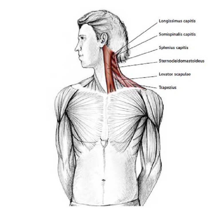
Diagram of the shoulder, including the location of the rotator cuff. The following is an overview of the shoulder muscle anatomy. Related posts of diagram of shoulder muscles and tendons muscle anatomy fitness. Postural and active movement muscle, used to tilt and turn the head and neck, shrug, steady the shoulders, and twist the arms. It also helps you raise and rotate your arm. The rotator cuff is a collection of muscles and tendons that surround the shoulder, giving it support and allowing a wide range of motion. What are common rotator cuff injuries? The main shoulder muscles are trapezius, deltoid, pectoralis major and 4 rotator cuff muscles: Deltoids anatomy when most people think of the The muscles of the shoulder bridge the transitions from the torso into the head/neck area and into the upper extremities of the arms and hands. The scapulae, posteriorly, the clavicles anteriorly and completed anteriorly by the manubrium of the sternum (part of the axial skeleton). While seated, have your partner place one hand at the front of your shoulder joint and one hand at the rear. Superficial muscles are the muscles closest to the skin surface and can usually be seen while a body is performing actions.
Related posts of diagram of shoulder muscles and tendons muscle anatomy fitness. See more ideas about shoulder anatomy, anatomy, muscle anatomy. The muscles in the shoulder aid in a wide range of movement and help protect and maintain the main shoulder joint, known as the glenohumeral joint. The partner should slowly, but firmly press on both sides of your shoulder to compress the ac joint. The following is an overview of the shoulder muscle anatomy.
Labeled Anatomy Chart Of Neck And Shoulder Muscles On ... from media.istockphoto.com The main shoulder muscles are trapezius, deltoid, pectoralis major and 4 rotator cuff muscles: Ebraheim's educational animated video describes muscle anatomy of the shoulder girdle and anatomy of the shoulder joint.anatomy of the shoulder muscles a. It is a flat, gliding joint. The muscle elevates, depresses, rotates, and retracts the scapula, or shoulder blade. The shoulder blade (scapula) connects to the collarbone (clavicle) at this joint. As the name implies, the rotator cuff functions to allow you to rotate your shoulder and lift your arm. The muscles of the shoulder support and produce the movements of the shoulder girdle.they attach the appendicular skeleton of the upper limb to the axial skeleton of the trunk. Around the shoulder, muscles in the back, neck, shoulder, chest and upper arm all work together to support and move the shoulder.
The deltoid is the largest, strongest muscle of the shoulder.
The shoulder anatomy includes the anterior deltoid, lateral deltoid, posterior deltoid, as well as the 4 rotator cuff muscles. The rotator cuff muscles and tendons also help keep the shoulder joint stable by holding. While seated, have your partner place one hand at the front of your shoulder joint and one hand at the rear. Muscle structure of the knee 12 photos of the muscle structure of the knee muscle anatomy knee mri, muscle anatomy of the knee, muscle anatomy of the knee joint, muscle and tendon structure of the knee, muscle structure of the human knee, human muscles. Muscles allow us to move by pulling on bones. The shoulder is a complex combination of bones and joints where many muscles act to provide the widest range of motion of any part of the body. The bursa is a small sac of fluid that cushions and. This flexibility is also what makes the shoulder prone to instability and injury. The shoulder blade (scapula) connects to the collarbone (clavicle) at this joint. Deltoids anatomy when most people think of the Around the shoulder, muscles in the back, neck, shoulder, chest and upper arm all work together to support and move the shoulder. The shoulder muscles are responsible for maintaining the widest range of motion of any joint in your body. Subscapularis, supraspinatus, infraspinatus and teres minor.